Who has an OpenSPIM
Feel free to edit this page with information about your system! (You will need a GitHub account)
Aachen, Germany

OpenSPIM with temperature controlled sample chamber by Fraunhofer IPT
- Life Sciences Engineering Business Unit at the Fraunhofer Institute for Production Technology IPT.
Our OpenSPIM design has a temperature controlled sample chamber for long-term experiments and uses a timing belt for precise sample rotation.
Dresden, Germany
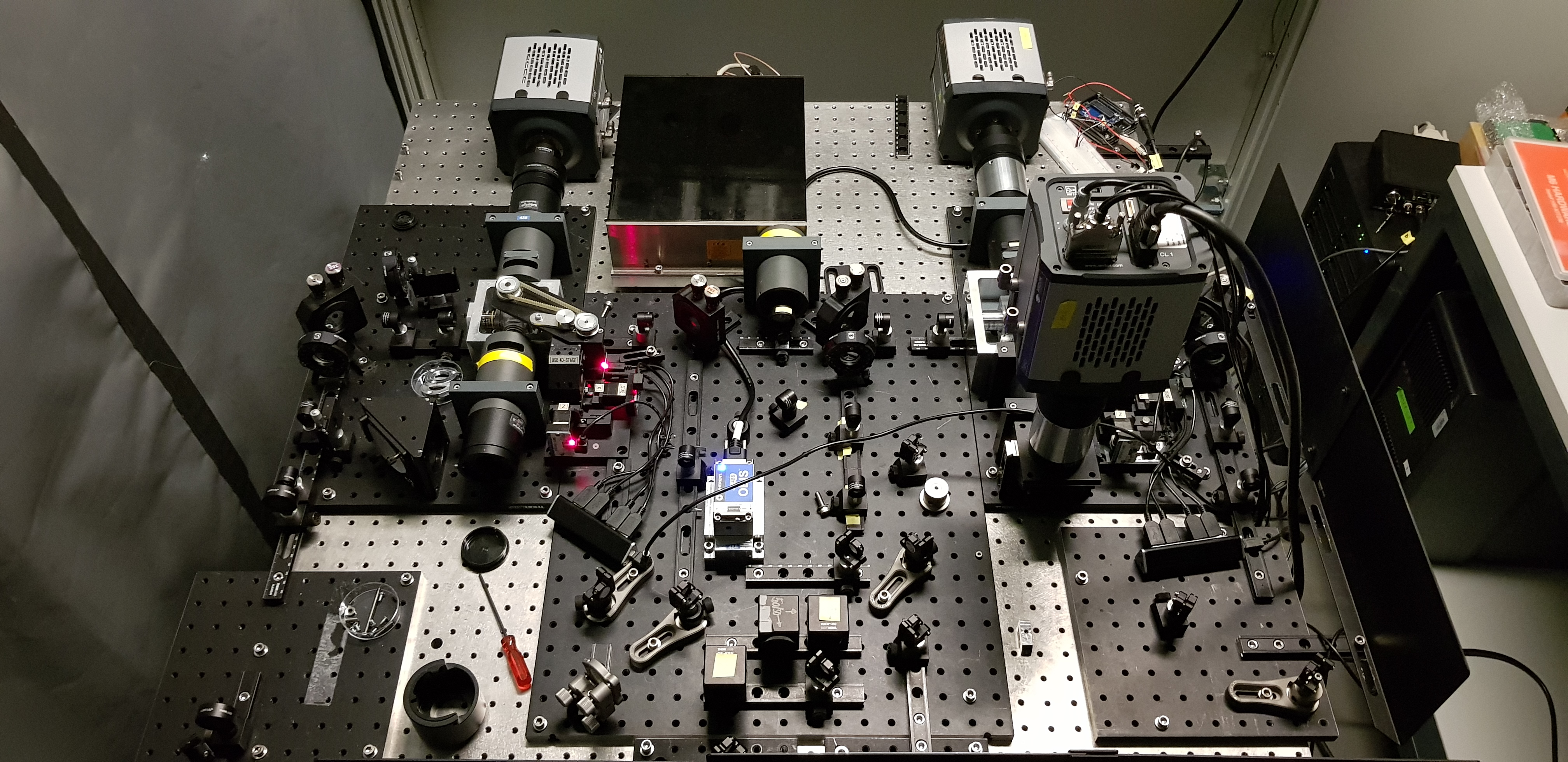
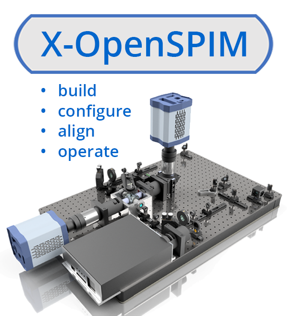
Tomancak Lab X-OpenSPIM
Tomancak group at the Max-Planck Institute of molecular Cell Biology and Genetics.
- X-OpenSPIM sample chambers (40x Nikon detection objectives) for dual-sided illumination and dual-sided detection
- T-OpenSPIM sample chambers (20x, Olympus detection objective) for dual-sided illumination and single-sided detection
- Andor sCMOS Neo cameras
- 488/561 VersaLase (X-OpenSPIM)
- 488 Coherent OBIS Laser (T-OpenSPIM)
- hardware-controlled laser triggering (ArduinoUNO)
- operated by MicroManager using our new custom made µManager plugin called µOpenSPIM
For details on how to assemble, configure, align and operate the X-OpenSPIM take a look at the openspim.org X-OpenSPIM website and read our related publication.
Madison, WI, USA
LOCI's L-SPIM, Jan. 22, 2016
The Laboratory of Optical and Computational Instrumentation (LOCI) run by Kevin Eliceiri at the University Wisconsin-Madison. It uses a Coherent Cube laser and a Hamamatsu Orca R^2 camera, being controlled by a 32-bit Windows XP computer.
The OpenSPIM software was developed in this lab by Johannes Schindelin and Luke Stuyvenberg, and tested and refined with the help of Julie Last and Jayne Squirrel (see the People page for details), using that setup. The source code is hosted on GitHub.
Heidelberg, Germany
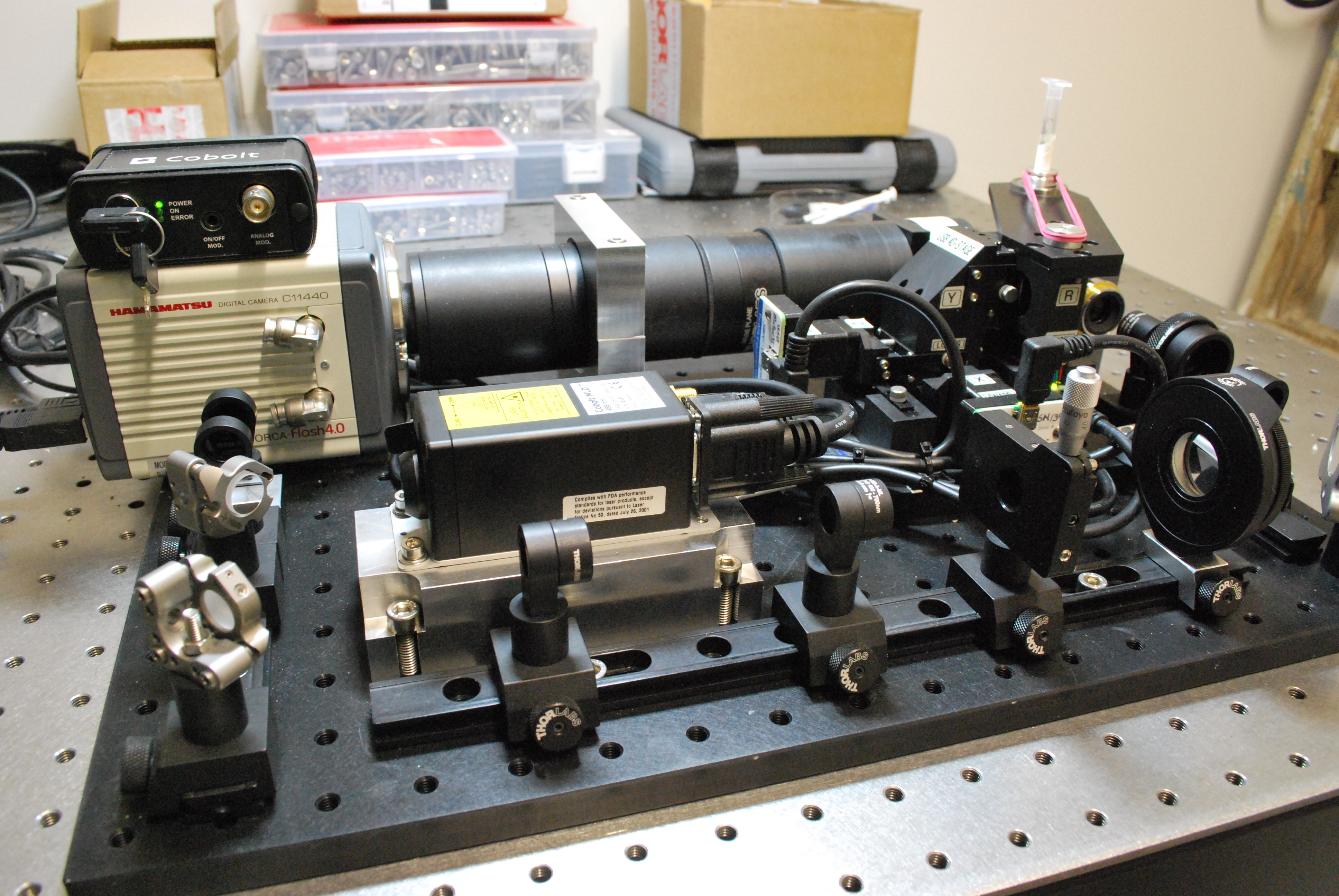
COS Heidelberg OpenSPIM
Maizel and Lemke groups at the Center for Organismal Studies of Heidelberg University.
This setup slightly differs from the specifications of OpenSPIM v1.0. In particular we use a 488 nm Cobolt laser, Nikon lenses and an Orca-Flash4.0, the whole running under Windows7 Pro.
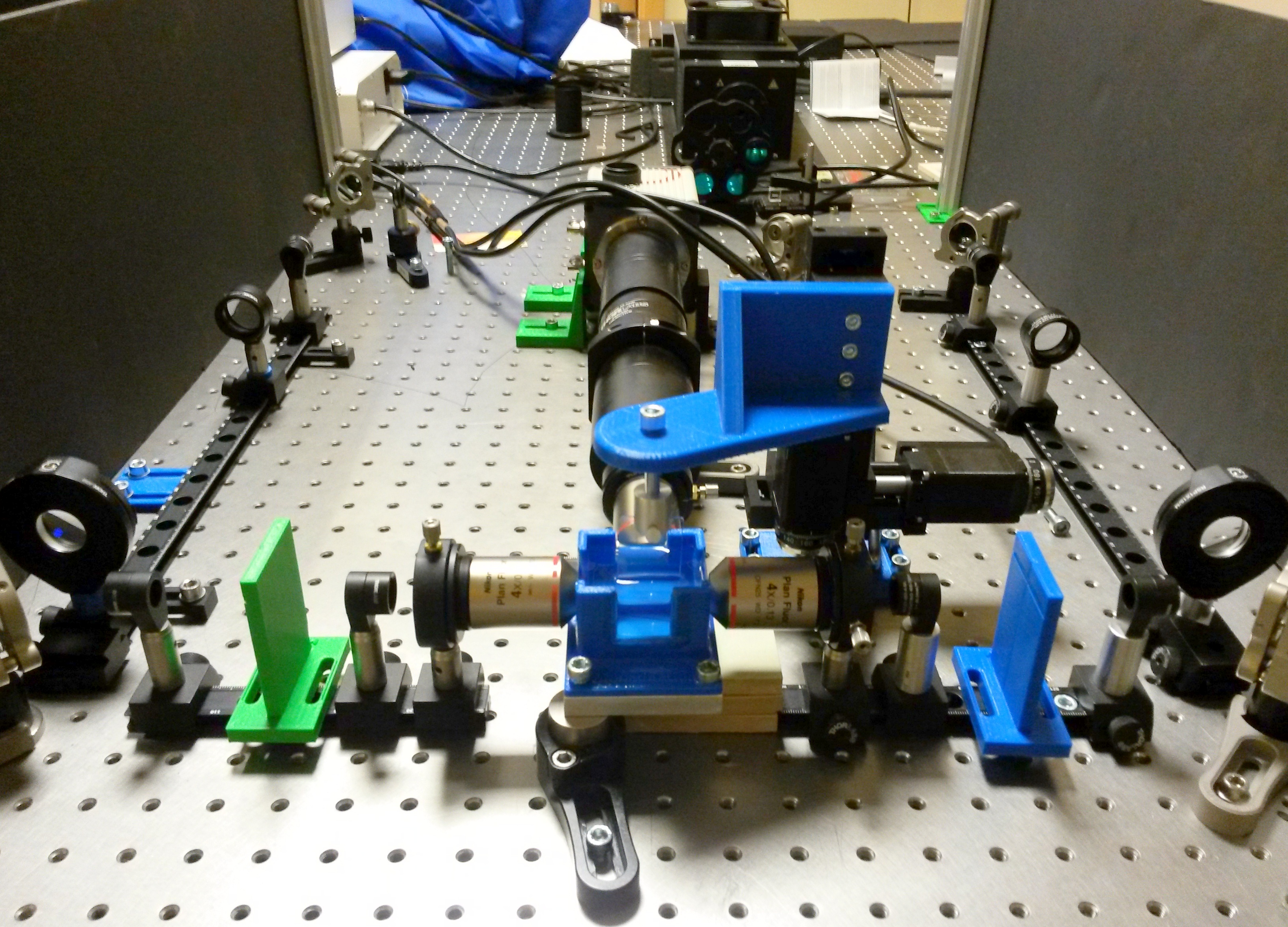
Cambridge, UK, Maths Department
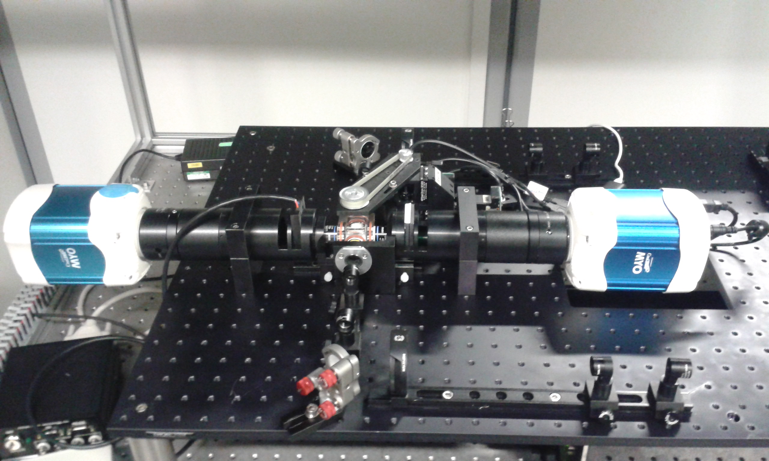
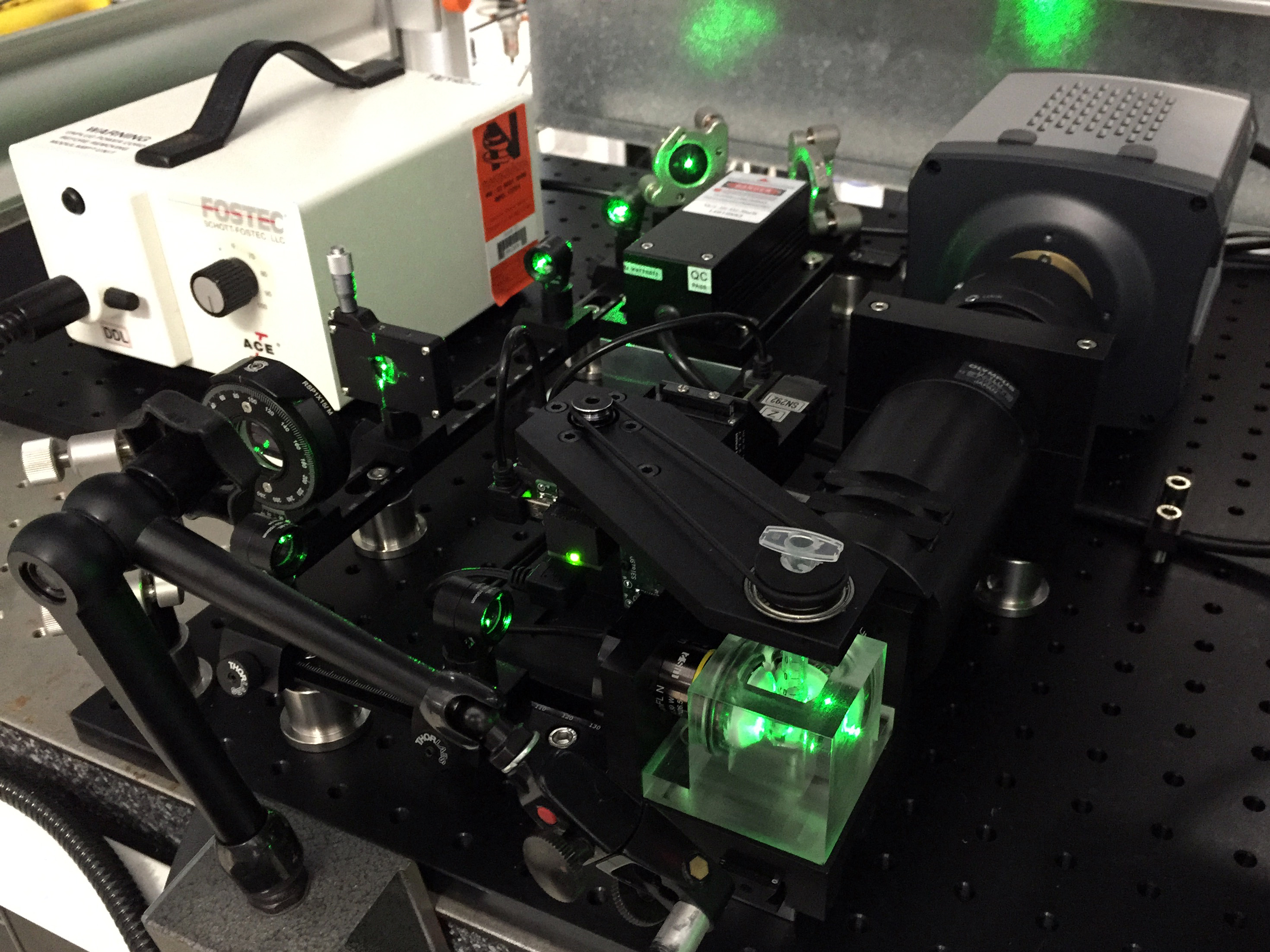
Fig. 1 Four-arm OpenSPIM
The Goldstein group in the Department of Applied Mathematics and Theoretical Physics at the University of Cambridge.
We recently extended to a 4-armed system (Fig. 1):
-
dual sided illumination with Vortran VersaLase multiple wavelength laser module (488, 561, 647 nm).
-
dual sided detection with 2 Photometrics Coolsnap Myo.
-
We included a Thorlabs shutter in the beam path.
-
For brightfield illumination during sample positioning we use a lamp that is temporarily inserted in one of the filter holders (Fig. 2)
As part of the Biomaker Challenge 2018 we added the following modifications:
-
Adjustable metal feet were attached to the clamps that carry the detection arms to align the two cameras (Fig. 3)
-
We wanted to provide illumination for light dependent (photosynthetic) organisms between acquisitions of a time lapse recording.
For this we added a lamp that switches on when the laser shutter closes. The LED driver is triggered by the laser shutter controller via a TTL inverter (Fig. 4).
- To improve the robustness of the sample positioning we designed 3D printable sample holders (Fig. 5).
See also Dual-view imaging on hackster.io
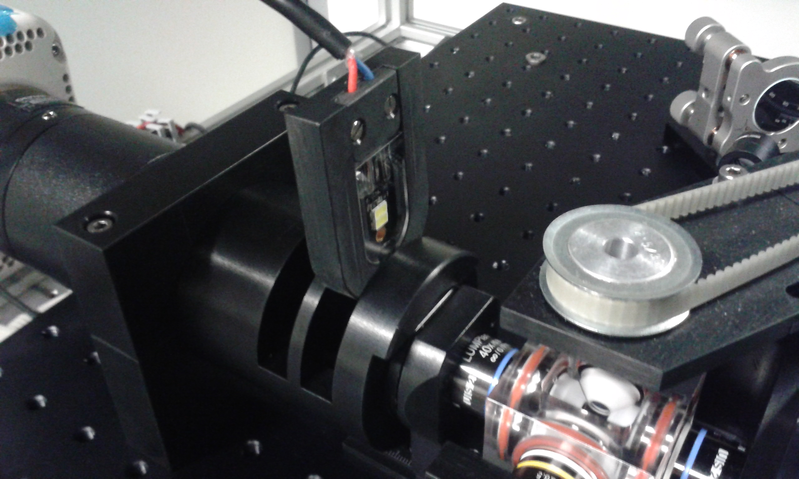
Fig. 2 LED tube light for brightfield
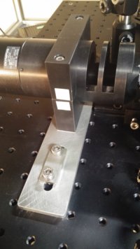
Fig. 3 Adjustable foot to align detection arm
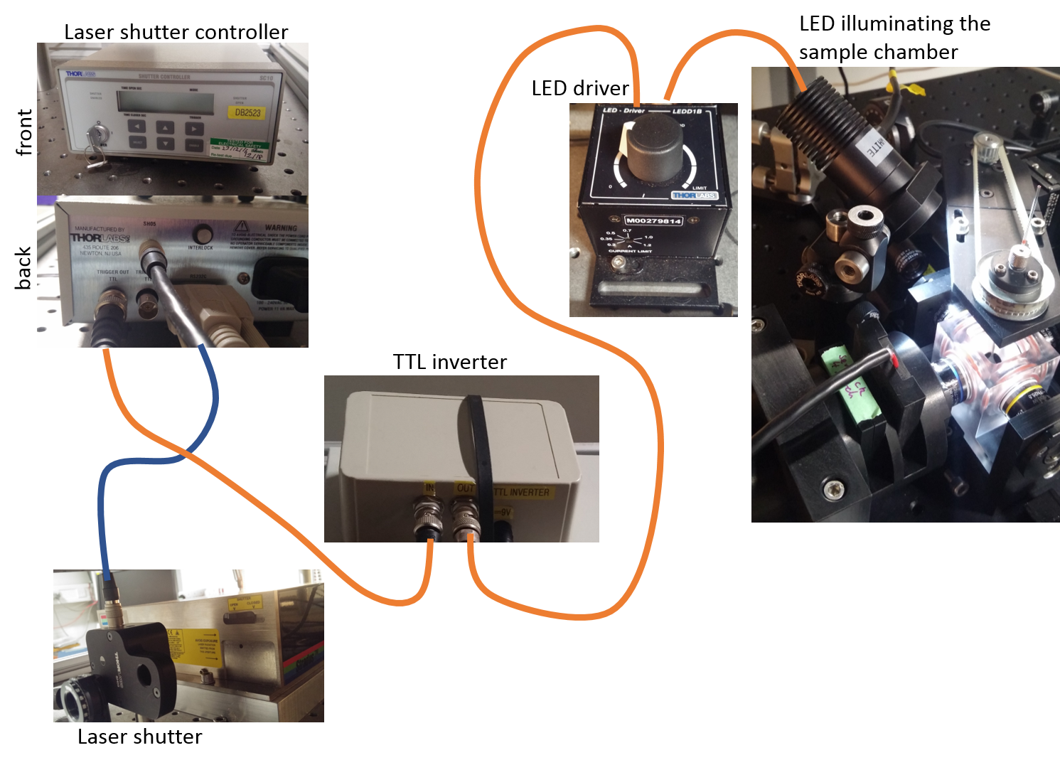
Fig. 4 Schematic for illumination between acquisitions
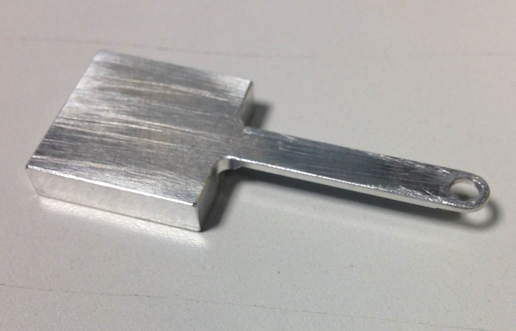
Fig. 5 3D-printed sample holder
Cambridge, UK, MRC LMB
The Light Microscopy Facility at the Medical Research Council's Laboratory of Molecular Biology
-
Our system features two-sided excitation with adjustable focus of the sheet lenses (Olympus 5x/0.15 air MPLFLN5X) which sit outside the imaging chamber to cope with different refractive indices. - 4 laser lines - combined in a separate laser bed and fed into a fibre (405nm & 640nm: both Coherent Cube; 488nm & 561nm: both Coherent Sapphire)
-
The imaging chamber can be heated to e.g. 37C for live cell imaging purposes. - Imaging lens is a Nikon 16x/0.8NA (N16XLWD-PF) with 3mm WD.
-
In front of the camera is a Thorlabs 6-position filter wheel (FW102C)
-
The entire system is portable (sort of) and modified to be laser safe as it is operated within our multi-user facility.
Melbourne, Australia
OpenSPIM system using the Picard high-resolution stage and an Andor Neo camera in the Petrou Lab, Melbourne, Australia.
The Petrou group at the Florey Institute, the Centre for Neural Engineering and the Centre for Integrated Brain Function at the University of Melbourne.
- We are deploying OpenSPIM for CLARITY and Scaleview based imaging of brain tissue for studying neural morphology. We use Dragon Lasers and an Andor Neo sCMOS camera.
Berlin, Germany
OpenSPIM at Humboldt University Berlin
The Scholtz group at the Humboldt University Berlin
-
I am interested in morphogenetic processes during early development of arthropods like the water flea Daphnia magna, the woodlouse Porcellio scaber or the beachhopper Parhyale hawaiensis. Using the SPIM technique for live imaging experiments enables us to observe dynamic and complex processes like gastrulation and beginning morphogenesis.
-
We implemented a Coherent Obis 561 nm laser and a Hamamatsu OrcaFlash4 sCMOS camera into the OpenSPIM system. The system runs at the moment with just one-side illumination. However, the sample chamber is already made for dual-side illumination (2x 10x illumination and 16x detection). Additionally we integrated a Nikon-module that allows us to increases our range of magnification (1x, 1,25x, 1,5x, 2x).

T-SPIM chamber, designed by Pete Pitrone and build at the workshop of the MPI CBG Dresden.
Exeter, UK
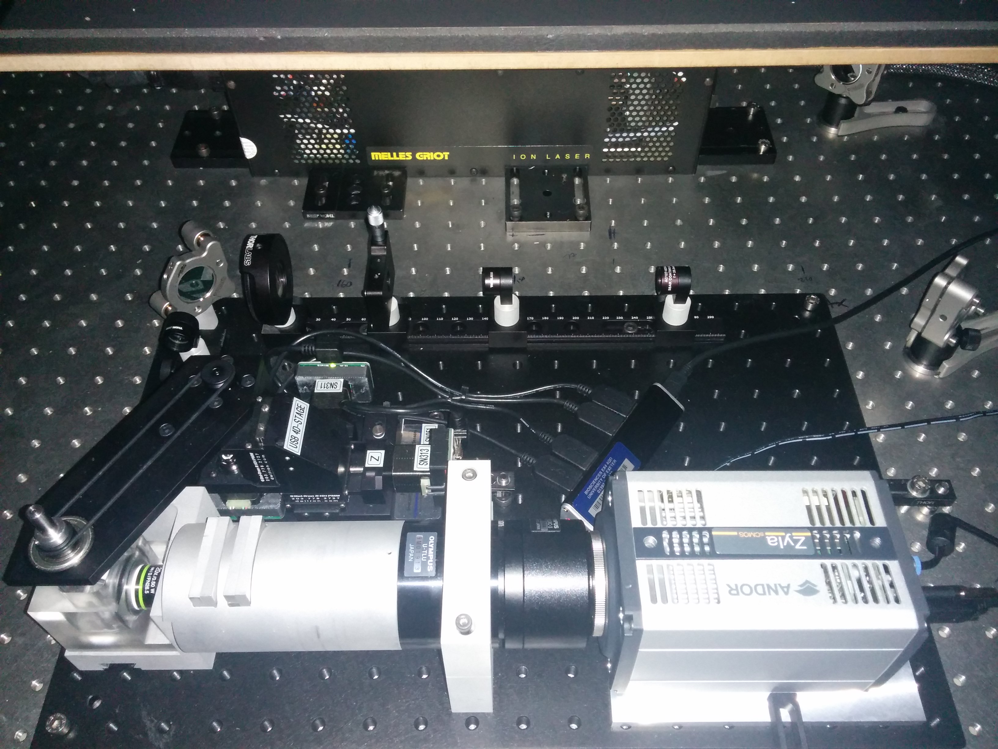
OpenSPIM at the University of Exeter.
The College of Life and Environmental Sciences in association with the College of Engineering, Mathematics and Physical Sciences.
-
We have constructed the system with a Melles Griot 488nm Argon laser and a Zyla 5.5 sCMOS camera running on 64-bit Windows 7 to image transgenic zebrafish.
-
The system currently has magnification options of 0.5x and 1x and 1-photon illumination with plans to upgrade to 2-photon in the future.
Oulu, Finland
OpenSPIM at the University of Oulu.
The Light Microscopy Core Facility, at the Biocenter Oulu and the University of Oulu.
-
Modified T-SPIM: dual side illumination with Vortran Stradus 488nm and 561nm lasers, ScopeLed G180 for bright field/transmitted light imaging, Olympus UMPLFLN 20X/0.5 W and Olympus LUMPLFLN 40X/0.8 W detection objectives, Hamamatsu ORCA-Flash 4.0 V2 camera. - Temperature, CO2 and O2 controlled liquid exchange/perfusion system for live cell imaging.
-
OpenSPIM has been used for live cell imaging: different cell types and spheroids in different 3D culture matrices, e.g. in Matrigel, collagen, myogel and fibrin mounted inside FEP tubes, and also for imaging living mouse skin cancer biopsies. It has also been used for whole mount stained and/or transgenic (expressing tdTomato, GFP, mCherry…) fixed samples, typically mouse embryonic kidneys, pieces of adult mouse tissues like heart, liver, fat, lung etc.
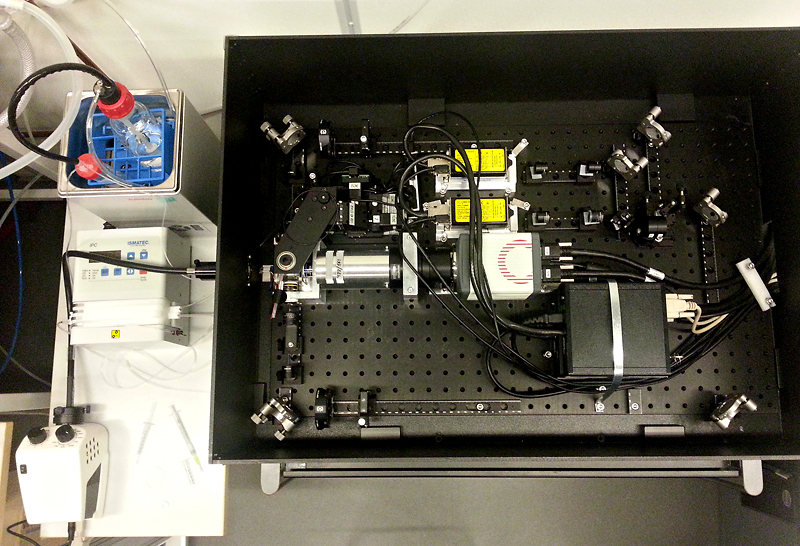
Indianapolis, IN, USA
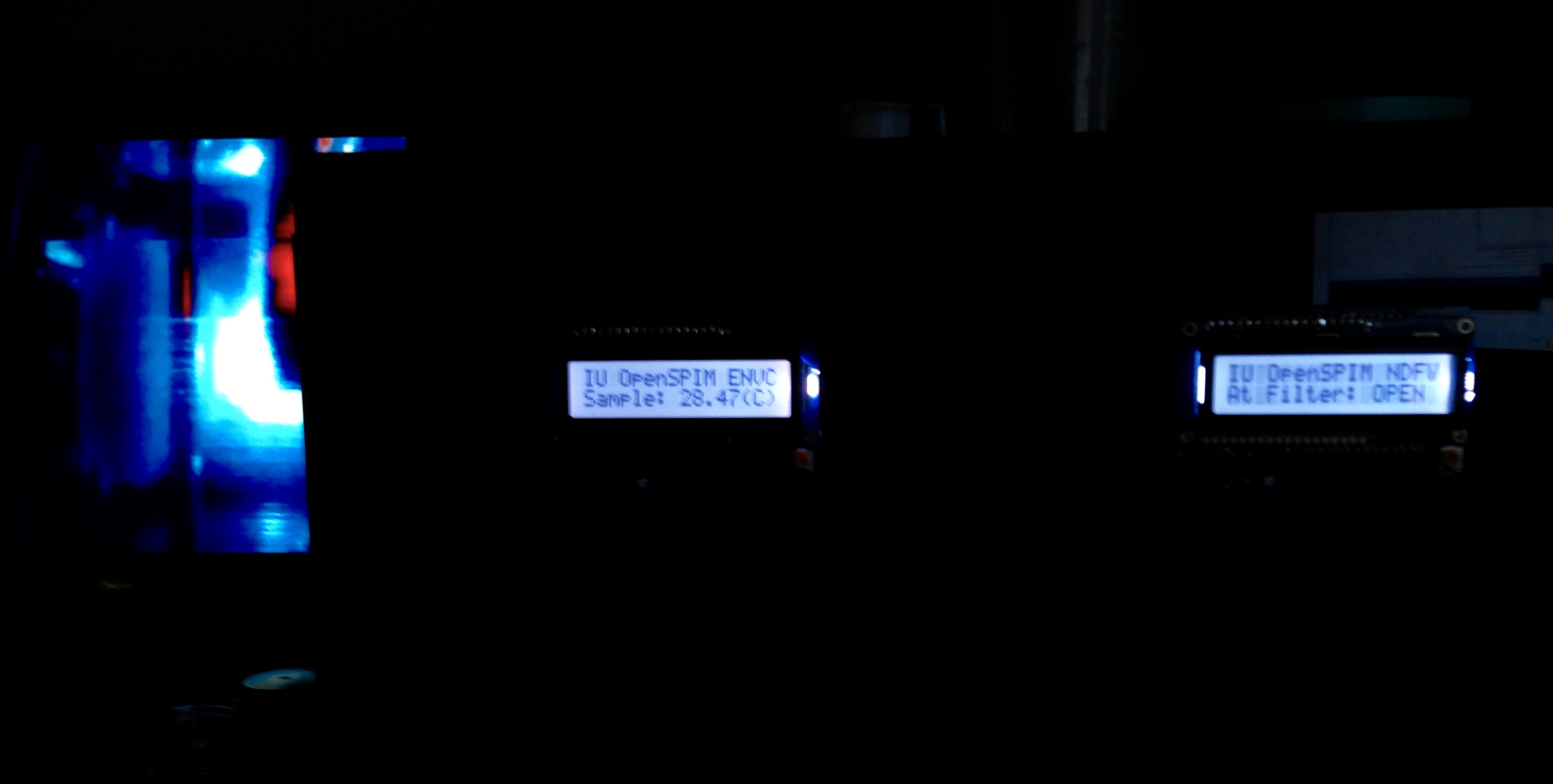
OpenSPIM at IUSM.
OpenSPIM chamber with oblique illumination.
The Indiana Center for Biological Microscopy at Indiana University School of Medicine.
-
We extended the base L-SPIM design for use in a core facility by including additional excitation wavelengths and support for live imaging. We are imaging with an ORCA Flash 4.0 v2 using CameraLink.
-
To support more fluorophores we incorporated five laser lines (442, 488, 514, 568 and 647 nm) with AOTF modulation controlled via an Arduino based 6 channel DAC. Emission wavelength filtering is with a workhorse Sutter Lambda 10-2. Multichannel acquisition is fully controlled (AOTF, filter wheels, etc) by a custom version of the SPIM Acquisition Plugin and derivations of the Arduino device adapter for MicroManager. To assess laser power, a motorized mirror and ThorLabs detector have been incorporated prior to beam shaping.
-
To support live imaging we have incorporated a perfusion and temperature control system into the standard L-SPIM chamber(no drilling etc.) built with off-the-shelf components (shout-out to Adafruit and Sparkfun). It is Arduino based and relies on a bunch of pumps and thermistors. MicroManager integration and a CO2 loop is in the works (anyone with experience implementing amphenol CO2 sensors?).
-
Our OpenSPIM is fully contained to simplify laser safety. To facilitate alignment and sample positioning a secondary camera driven by a RaspberryPi provides a live image of the chamber with oblique RGB LED illumination from an Arduino and PWM via the 16 channel TLC5940.
-
To aid in sample positioning a knob bar is under development with four free-turning knobs-one for each axis with position feed-back. - IU OpenSPIM has been used for imaging of Zebrafish (live heart-beat and fixed and cleared tissue), organoids, and tissue samples including rat kidneys-most in FEP tubing. It has also been a testbed for new technologies and methodologies.
Whole-brain imaging based on OpenSPIM, Warsaw, Poland
Cleared-tissue imaging SPIM at the Nencki Institute.
We are the Laboratory of Neurobiology at the Nencki Institute of Experimental Biology, Warsaw, Poland
-
Using OpenSPIM as the starting point, we developed a cleared-tissue imaging SPIM system using dry 4X/0.13 Nikon objectives with WD of 17 mm, or about 25 mm in immersion
-
We use 3D printed chambers for CLARITY- and CUBIC-cleared tissue and glass cuvettes for BABB-cleared tissue
-
Recently we replaced the Picard stage with one by Standa, with greatly increased speed and travel range at comparable cost
-
Also recently a second illumination arm was added, converting the setup from the L to the T configuration
-
Ar-Ion laser (458/488/515 nm with manual filter wheel for wavelength selection) used for illumination
-
Beam height is raised to 65 mm, mainly for compatibility with the argon laser
-
We have demonstrated whole mouse and rat brain imaging (paper)
Newcastle (HMRI/UoN), Australia
OpenSPIM HMRI/UoN glamour shot
The 3D Tissue Clearing and Lightsheet Microscopy Facility (HMRI/UoN).
-
Our focus is on the development and progression of 3D histology in medical and plant sciences.
-
Imaging is performed with an OpenSPIM-based T-SPIM with Hammamatsu Orca Flash, Arduino controlled 405, 473 and 532nm CNI lasers, and Thorlabs emission filters and dichroics
-
Picard 4D stage controlled by wireless 3D 'spacemouse' - highly recommended.
-
Controlled via Micromanager, with 3D data processing performed with FIJI, Terastitcher and Vaa3D using the UoN HPC Cluster
-
Currently building a new SPIM incorporating a digitally scanned lightsheet (to take advantage of the lightsheet mode of the Orca Flash), two Nikon x4 Plan Fluorite illumination objectives (17mm WD), 1" optics, enlarged chamber, and a long working distance RI corrected detection objective for imaging large, cleared tissues
London, UK
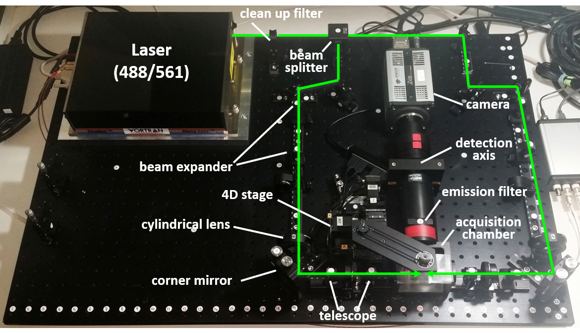
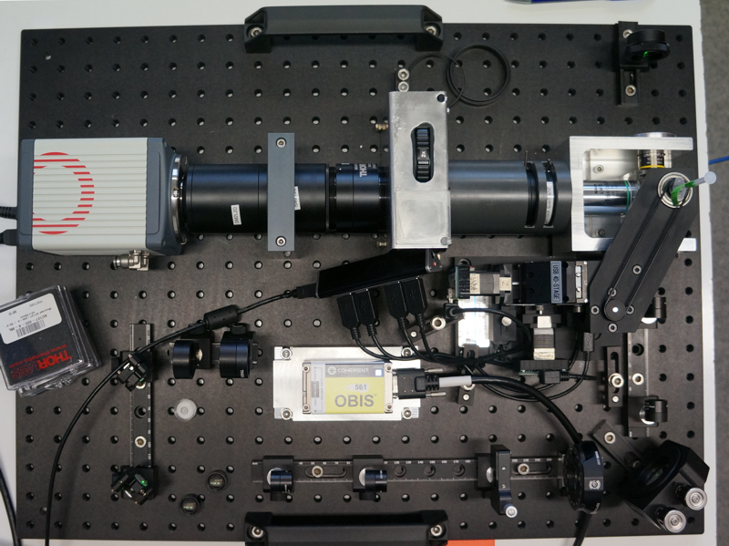
OpenSPIM with two lasers (VersaLase: 488, 561) and dual sided illumination running in the the Max Telford Lab (UCL, UK)
OpenSPIM running at the Max Telford Lab, University College London
-
It builds on the standard design with the addition of two colour laser illumination (VersaLase™ Multiple Wavelength System) and dual sided illumination.
-
To reduce image acquisition time ESio’s TTL controller box was incorporated
-
The OpenSPIM is used for high resolution 3d images and time lapse recordings to study the development of different lophotrochozoan members (Spiralia) in particular the polyclad flatworm Maritigrella crozieri.
-
For detailed information of our system and our experience of building it you can read our paper: Light-sheet microscopy for everyone? Experience of building an OpenSPIM to study flatworm development (BMC Developmental Biology, 2016)
Oldenburg, Germany
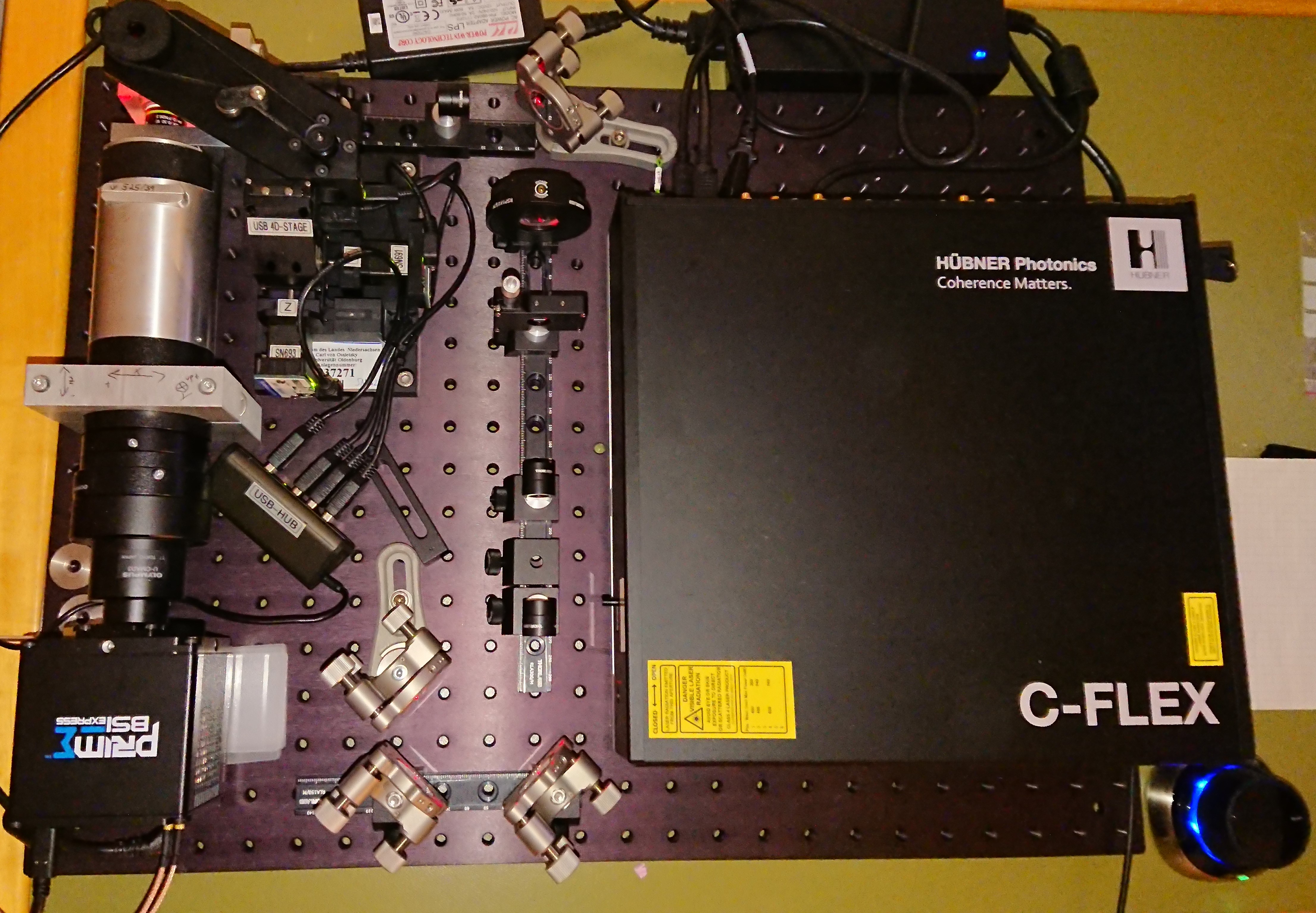
OpenSPIM at IBU Oldenburg.
Our OPENSPIM is located in the Sensory Biology of Animals group of Prof. Winklhofer in the Department of Biology and Environmental Science (IBU) at the University of Oldenburg:
https://uol.de/en/biology/groups-our-research/sensory-biology-of-animals
Component details:
-
C-FLEX C6 Laser combined with 405nm, 488nm and 633nm lasers (upgradable to 6 different laser lines)
-
Camera: BSI- Prime Express sCMOS
-
Sample chambers: Immersed chamber and dry chamber
-
Adapted illumination path for better compatibility with the C-Flex box
-
The entire set-up is mobile
-
3-D mouse
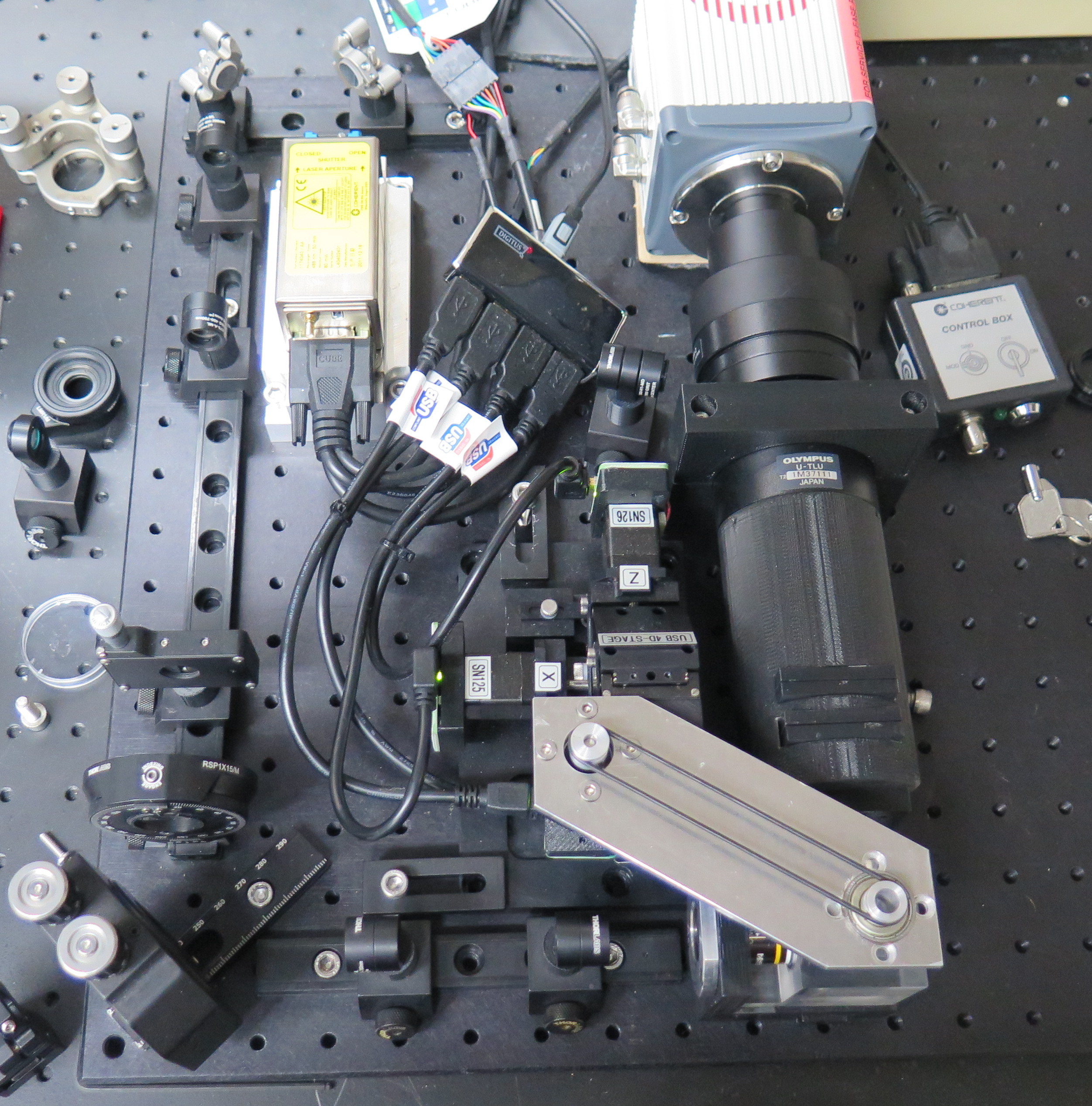
OsloSPIM at the University of Oslo, Norway
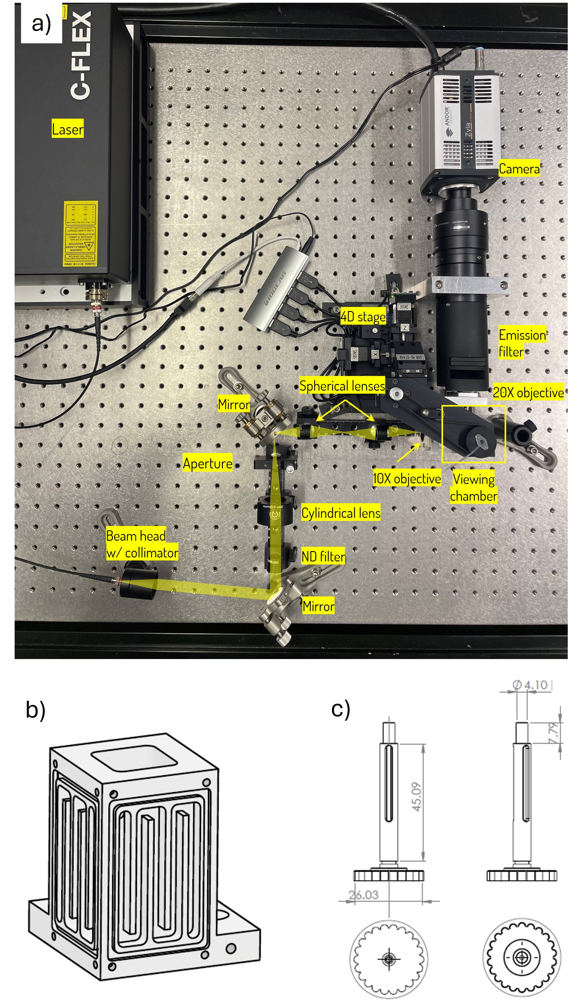
a) Schematic overview of the OsloSPIM setup. b) Water at physiological temperature flows through channels in the viewing chamber. c) The sample holder has gears to ensure slip free rotation. CAD-files repository: https://doi.org/10.17605/OSF.IO/GRK2W
We use our OpenSPIM to study how early mammalian embryo models (using so-called mouse gastruloids) grow and develop, and how the first organs form. Especially, we are interested in how cells self-organize and orchestrate their motion to form tissue. To explain the underpinning dynamics, we couple our experiments with 3D simulations. To fully utilize light-sheet imaging, we also study collective cancer cell migration and how nerve cells make new connections in live zebrafish, and we study the cells' own recycling system (autophagy) in Drosophila larvae.
https://www.uio.no/ritmo/english/people/postdoctoral-fellows/endrejm/index/
-
The illumination laser (Cobolt 6 series, Hübner Photonics) has laser heads with four different wavelengths (λ = 375, 488 561, 647 nm), corresponding to UV, green, red and far red light.
-
Fast imaging: An Arduino UNO board sends trigger pulses to the laser to obtain fast switching between the different wavelengths (ideal for live imaging).
-
Micro Manager controls the laser, camera (Andor Zyla 5.5, Andor), moveable 4D stage (USB-4D-Stage, Picard Industries), and the Arduino UNO board.
-
Temperature control: To control the temperature, water at physiological temperature flows through channels in the viewing chamber, which is made of heat conducting aluminum.
-
Media exchange: To provide fresh oxygen and nutrients to the 3D cell cultures, the media can be exchanged continually.