Optical Tissue Clearing
This page concerns optical tissue clearing (that is, rendering it transparent in order to image deeper) and imaging in a setup based on OpenSPIM.
Tissue clearing
There now exist a range of tissue clearing techniques for both mammalian and plant tissues. From imaging point of view, they fall into two categories:
-
solvent-based methods (BABB, ...DISCO) result in refractive index in the 1.55-1.56 range. These methods usually lead to sample shrinkage. The solvents are incompatible with most objectives and materials.
-
hydrogel and other aqueous methods (CLARITY, Scale, CUBIC etc. ) yield samples with refractive index 1.47-1.49. Samples often swell. Ordinary immersion objectives can be used.
Online resources for tissue clearing protocols
-
For a summary of various clearing techniques, and detailed instructions for performing CLARITY (excellent for neural tissue), please consult the manual compiled by the 3D Tissue Clearing and Lightsheet Microscopy Facility (HMRI/UoN)
Immersion media
Generally cleared tissue has a higher refractive index than water. To prevent losing focus when moving the sample, you have to use immersion medium with the same RI as the sample.
Most protocols recommend using the same reagent for RI matching and for imaging. This has some disadvantages - some reagents are too expensive to fill the entire light-sheet microscope chamber, others sometimes crystallize, react with outside air etc. Cheap and user-friendly alternatives for imaging include
- glycerol in the right concentration (80% has been used for CLARITY by Epp et al.)
- microscopy immersion oil
- the authors of CUBIC paper recommend a a 1:1 mixture of silicon oil TSF4300 (Momentive Performance Materials, RI = 1.498) and mineral oil (Sigma-Aldrich, RI = 1.467).
Objectives & chambers
Optimally, an immersion objective matched to the tissue refractive index should be used. Such objectives exist (by Zeiss, Olympus and Leica at the time of writing), but are very expensive. Long working distance air and water objectives can be used as well, but their performance will be somewhat limited by aberrations. Keep in mind that working distance changes if the immersion medium RI changes - this has to be taken into account when designing the setup.
Sample mounting
For confocal or two-photon microscopy, a chamber with depth equal to the sample thickness should be be used. For designs see:
For SPIM, the sample had to be suspended in a chamber filled with immersion medium. One way to do it is glue the sample to a glass slide mounted in a holder that in turn is mounted in the SPIM setup in place of the capillary. To glue a cleared sample, first pat with lint-free tissue. Cyanoacrylic glues (like Superglue) tend to work - sometimes it helps to try two or three brands, they differ in composition.
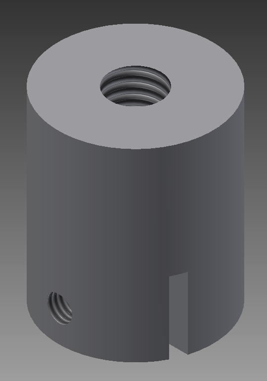
This holder can be mounted on the stage arm using a M6 screw. Glass slide can be mounted with a M3 nylon or nylon-tipped screw.