Early Zebrafish Development
Specification of Primordial Germ Cells (PGCs) in Zebrafish has 2 modes: one through maternal cytoplasmic inheritance of ‘germ plasm’ and this is common in most invertebrates and non-mammalians. The second is through induction of pluripotent epiblast cells during gastrulation and is common to mammals. The common denominator in most organisms is the migration of PGCs to genital ridge, further proliferation and differentiation.
Plan for the course
We have two transgenic lines. We want to investigate the earlier stages when this phenotype is caused, and we think light-sheet microscopy would be a great tool to study this, since other microscopes often require dechorionation which is quite difficult at these early stages and can easily harm the embryo.
-
PGC-EGFP: Marks the first PGCs (3-4hpf visible for the first time) - sample preparation described in Blaser et al. 2005
-
Buc-EGFP: Bucky Ball protein tagged with GFP. - sample preparation described in Bontems et al. 2009
Microscope description
We used four lens (water, 10x/0.3 objectives) SPIM setup in Jan Huisken lab. The details of SPIM setup and analysis are described in Schmid et al. 2013.
Migration of PGC-EGFP towards genital ridge
Tracking the migration
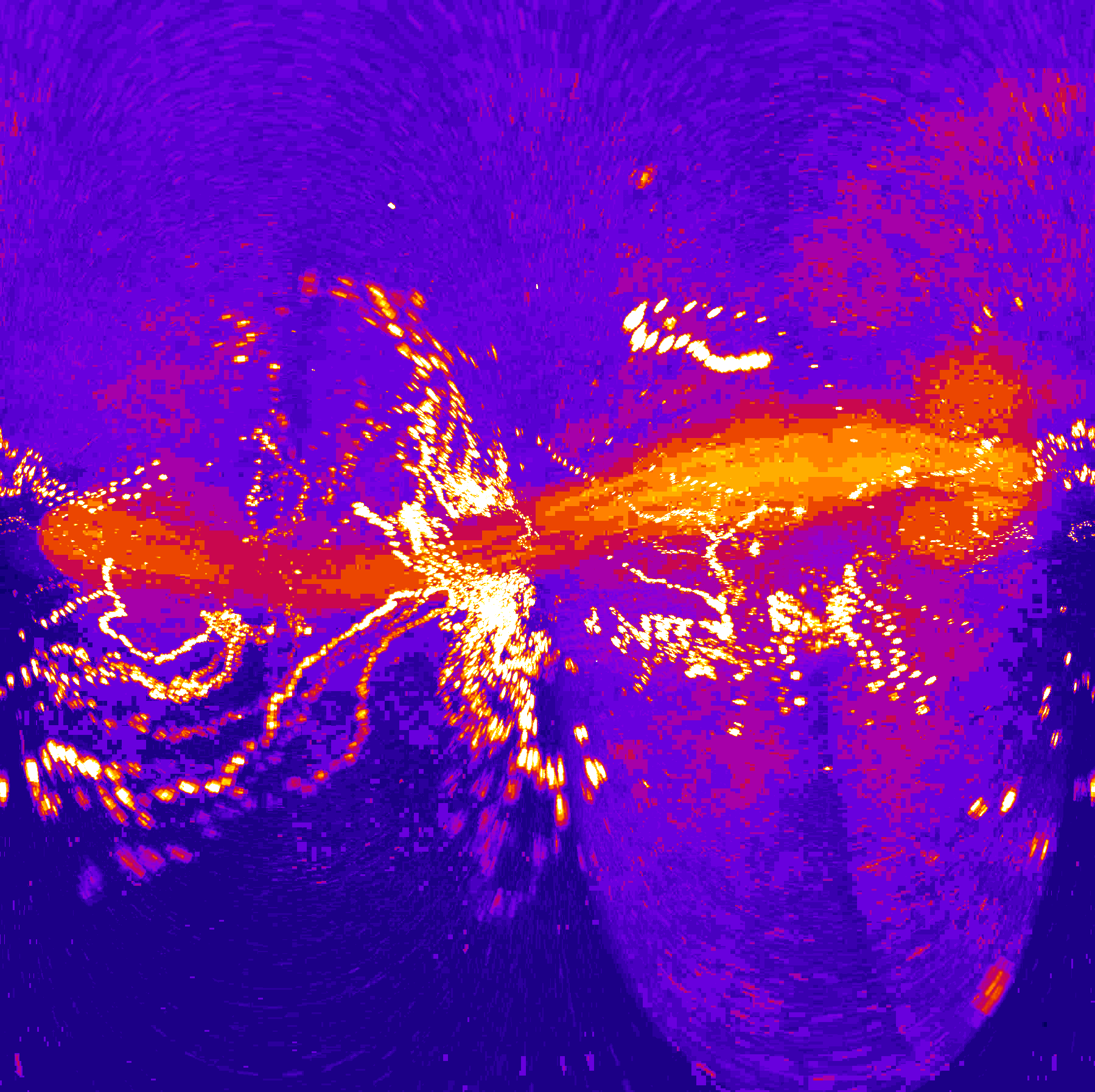
Maximum intensity projection.
Temporal color code.
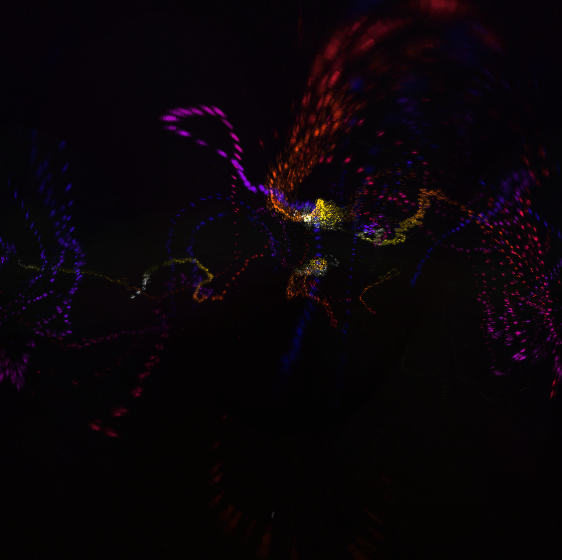
Localisation of Buc-EGFP to the cleavage planes
Buc is maternally deposited and this protein will be localizing to the cleavage planes directly after fertilization and can be imaged right after fertilization
Temporal color encoding of bucky ball movement
Acknowledgements
Thank you to Gopi Shah for teaching the basics of sample preparation, mounting and for the analysis.