SPIM for 3D tissue engineering imaging
In Tissue Engineering (TE) research field there is a need of 3D non-destructive imaging techniques capable of rendering images over time to investigate cells differentiation and proliferation; dynamics of the cell behaviors; cells interactions with each other and with the biomaterial; etc. In this project I want to answer to the questions: Can I use SPIM to image TE based in hydrogels? Can the samples be prepared inside FEP tubes? How deep I can image inside the gel without loosing resolution?
TE constructs of alginate with neurons
Rat primary cortical neurons derived and encapsulated in alginate hydrogel, maintained in culture for 5 days, stained with calcein AM (Invitrogen) and fixed in 4% formaline.
Samples fixed in FEP tubes with 0.5 mm
Image parameters:
- Dimensions (um): x- 487; y - 487; z - 341
- Objective: 10X, NA = 1
- Light Sheet thickness: 4.77 um
- Illumination wavelenght: 488 nm
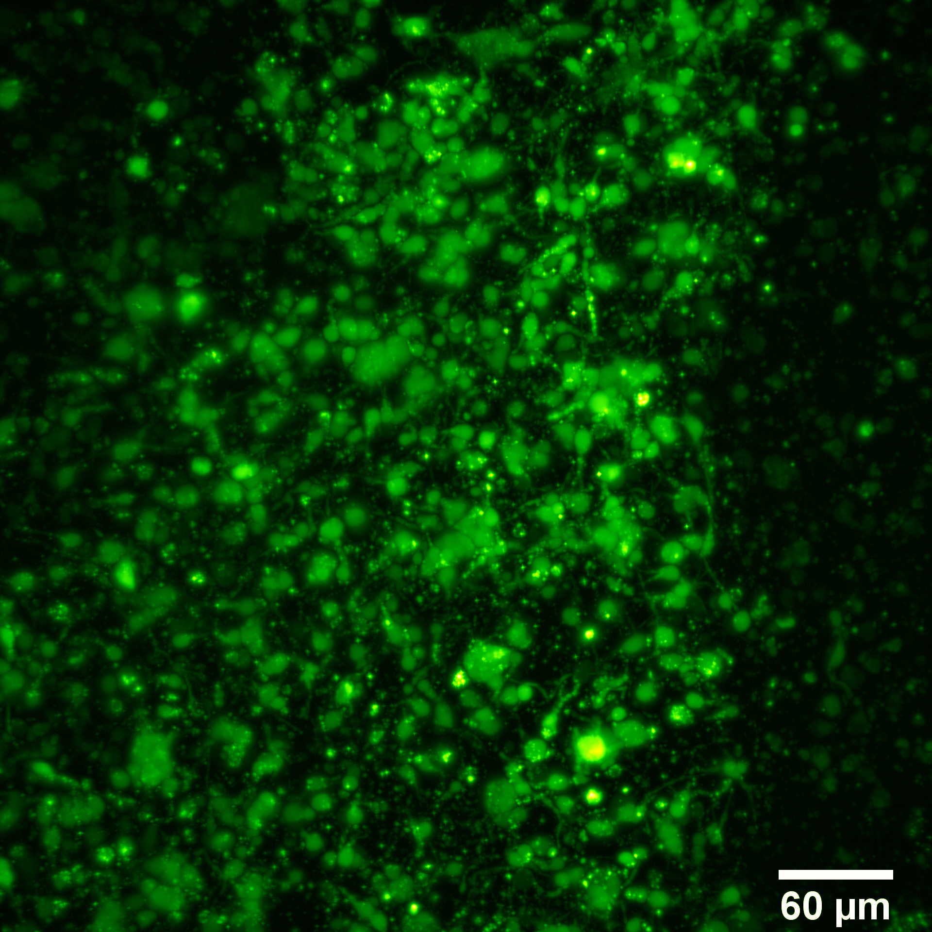
Maximum intensity projection
Video of the stack (video with better resolution here)
3D video (video with better resolution here)
Samples fixed in FEP tubes with 0.2 mm
Image parameters:
- Dimensions (um): x- 438; y - 438; z - 838
- Objective: 10X, NA = 1
- Light Sheet thickness: 4.52 um
- Illumination wavelenght: 488 nm
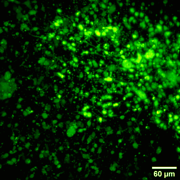
Maximum intensity projection
Video of the stack (video with better resolution here)
3D video (video with better resolution here)
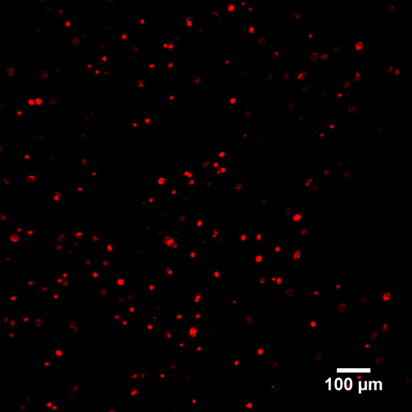
TE constructs of Gellan Gum with adipose stem cells
Sample fixed in FEP tubes with 0.5 mm
Image parameters:
- Dimensions (um): x- 552; y - 552; z - 59
- Objective: 10X, NA = 1
- Light Sheet thickness: 5.07 um
- Illumination wavelenght: 488 and 561 nm

Maximum intensity projection
3D video (video with better resolution here)
Sample imaged without the FEP tube
Image parameters:
- Dimensions (um): x- 1226; y - 1226; z - 731
- Objective: 10X, NA = 1
- Light Sheet thickness: 6.11 um
- Illumination wavelenght: 488 and 561 nm
Maximum intensity projection
3D video (video with better resolution here)
Conclusions
Although the TE constructs of alginate with neurons were not in good conditions (they were prepared 1 month before the imaging, and they were lost during the shipment for several days what resulted in the sedimentation of the neurons) and the stain used was not specific for the dendrites, the images look promising. From the images obtained, it can be clearly seen that better quality images were obtained with the thinner FEP tube. In the videos of the stacks we can also clearly see that there is a degradation of the images with the increasing of the depth (this videos start from the deepest image).
Concerning the TE constructs of Gellan Gum with adipose stem cells we can clearly see that the images are less blurry when the tube is not used, but Gellan Gum also affects the images.
In conclusion, SPIM is a promising technique to image TE constructs based in hydrogel. In order to get better image resolution I need to improve the sample fixation method.
Acknowledgements
I want to thank you to my collaborators that provide me the samples I imaged during this course, mainly, Gemma Palazzolo, Janne Koivisto and Jyrki Sivula. I also want to thank to the organizers and all the Instructors of the EMBO practical course.